Reducing patient stress during testing while improving the medical instrumentation used to detect cancer during checkups is truly a win-win for all involved. Such was the task facing researchers at Tokyo's Kitasato University's Center for Fundamental Sciences. The key focus of their research was to improve on traditional methods of cancer detection that do not provide sufficient resolution or r...
Reducing patient stress during testing while improving the medical instrumentation used to detect cancer during checkups is truly a win-win for all involved. Such was the task facing researchers at Tokyo’s Kitasato University’s Center for Fundamental Sciences. The key focus of their research was to improve on traditional methods of cancer detection that do not provide sufficient resolution or require the patient to undergo severe stress during the checkup.
With that goal in mind, Kitasato’s researchers set out to create a high-speed Fourier domain optical coherence tomography (OCT) system. OCT is a noninvasive imaging technique that provides subsurface, cross-sectional images of translucent or opaque materials. OCT images enable the visualization of tissues or other objects with resolution similar to that of some microscopes. In the academic community, there has been an increasing interest in OCT because it provides much greater resolution than other imaging techniques such as magnetic resonance imaging (MRI) or positron emission tomography (PET). Additionally, the method is extremely safe for the patient because of very low laser outputs and no need for ionizing radiation.
The 30.3 dB reflection is attenuated with neutral density filters. The thick solid curve is the noise floor measured with the sample light blocked. Sensitivity determined by these values is scaled at the right-hand side vertical scale.
OCT uses a low-power light source and the corresponding light reflections to create images—a method similar to ultrasound. The difference is that light is monitored instead of sound. When a beam is projected into a sample, much of the light is scattered, but a small amount reflects as a collimated beam, which can be detected and used to create an image.
Light into images
The Fourier domain OCT system developed by Kitasato’s researchers uses optical demultiplexers to enable separation of 256 narrow spectral bands from a broadband incident light in a 25.0 GHz frequency interval centered at 192.2 THz (1559.8 nm wavelength). Separation of the spectrum enabled simultaneous detection of all the bands using the 60 MS/s sample rate of the 256 high-speed analog-to-digital converter (ADC) channels incorporated in National Instruments’ PXI-5105 digitizers used for data acquisition.
The NI PXI-5105 was selected for its high-channel density of eight inputs per board, which enabled the 256 high-speed-channel system to maintain a small footprint.
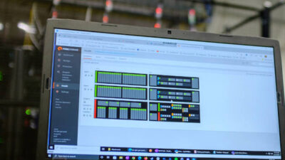
The system that was ultimately developed includes 32 of the eight-channel PXI-5105 digitizers distributed throughout three 18-slot NI PXI-1045 chassis. Digitizers in different chassis were synchronized using NI PXI-6652 timing and synchronization modules and NI-TClk synchronization technology, which provided phase coherency among channels in tens of picoseconds. LabView, National Instruments’ graphical programming application, was used for processing and visualization of the data.
Using the optical demultiplexers in a Fourier domain OCT system as spectral analyzers, OCT imaging of 60 million axial scans per second was achieved. Using a resonant scanner for lateral scan, 16 kHz frame rate, 1,400 A-lines per frame, and a 3 mm depth range, the system’s OCT imaging demonstrated a 23
System details
In the system developed at Kitasato, the light source is a broadband superluminescent diode (SLD, prototype by NTT Electronics). As shown in the schematic diagram graphic, the output light from the SLD is amplified with a semiconductor optical amplifier (SOA, COVEGA, BOA-1004 type) and divided equally into the sample arm and reference arm with the coupler (CP1). The output intensity of light from SOA1 is adjusted so that the power illuminating the sample is 9 mW, which conforms to the ANSI (American National Standards Institute) safety limit.
The OCT system then directs the sample arm light onto the sample (S) with a collimator lens (L1) and an objective lens (L2). A resonant scanner (RS, Electro-Optical Products, SC-30 type) and a galvano mirror (G, Cambridge Technology, 6210 type) are used to scan the light beam on the sample. Back-scattered or back-reflected light from the sample is collected by the OCT system’s light illuminating optics and directed to SOA2 (COVEGA, BOA 1004 type) with an optical circulator C1. The output of SOA2 and the reference light is combined with a coupler CP2 (50:50 coupling ratio). The reference arm comprises the optical circulator C2, the collimator lens L3, and reference mirror RM.
Outputs from CP2 are sent to two optical demultiplexers (OD1 and OD2) for balanced detection. The CP2 uses the same optical frequency as the two ODs with balanced photo-receivers (New Focus, 2117 type), which feature a total of 256 photoreceivers. The photoreceiver outputs are detected with the fast multichannel ADC system of 32 PXI-5105 digitizers, which store data in onboard memory during the single-shot acquisition before transferring it to a computer for analysis.
Distinct benefits
The OD (optical demultiplexers)-OCT is similar to SD (spectral domain)-OCT in that it detects the interference spectrum simultaneously. The difference is that the OD-OCT detects an entire interferogram at the speed of data acquisition at different frequencies simultaneously, instead of accumulating it into a CCD detector over a certain time period, as in the SD-OCT. Therefore, the system determines the axial scan rate by the data acquisition speed of the data acquisition system, which can be as fast as 60 MHz. The 16 KHz speed of the resonant scanner determines the frame rate.
Only one scanning direction is used for data acquisition (50% duty), which led to the sampling time of 31.25
Image shows physical setup of photoreceivers, demultiplexers, and data acquisition system linked to computer. Source: National Instruments
For this project, it was determined that the dynamic range of the system would be the ratio between the peak value of the point spread function (PSF) and the noise floor when the sample arm is not blocked. From test results, the dynamic range was estimated to be about 40 dB at all depths, slightly decreasing as the depth increases (see “Point Spread Function” graphic). A benefit of the OD-OCT is that the spectral width detected at each channel of the array wave guide is narrower than the frequency step of 25 GHz. A dynamic range of 40 dB is considered marginally sufficient for biological tissue measurements.
The penetration depth of the image is about 1 mm, which is shallow compared to the 2 mm penetration depth usually obtained with SS (swept source)-OCT or SD-OCT. This is due to the low sensitivity of the system. To render a 3D image, a number of OCT cross sections are required. Due to this issue and limited memory in the system, the sampling rate was reduced to 10 MHz.
Authors of this case study are: Dr. Kohji Ohbayashi, D. Choi, H. Hiro-Oka, H. Furukawa, R. Yoshimura, M. Nakanishi, and K. Shimizu, all of Kitasato University, Center for Fundamental Sciences. This application was named the overall winner in the 2008 Customer Application of the Year competition conducted during NIWeek 2008. Info at www.ni.com/niweek/best.htm


